
Peptide Mass Spectrometry Analysis: How Peptide Mapping Works
This guide explains how peptide mapping helps interpret mass spectrometry results and evaluate evidence for protein identification. It covers digestion, separation, fragmentation and database matching, showing how each stage contributes to the analysis.
It also outlines how these stages connect in a bottom-up workflow and what each can reveal. The goal is to help research teams distinguish the analytical question from the conclusions a result can support.
Key Takeaways
- Follow how enzymatic digestion and liquid chromatography prepare peptides for mass spectrometry, and see how each stage contributes to identification.
- Learn how fragmentation produces sequence evidence, including the b- and y-series ions commonly used in tandem analysis.
- Assess database matches as comparisons against reference sequences, rather than as de novo sequencing.
- Interpret peptide fingerprints within the full workflow, considering how modifications can affect measured masses and protein identification.
Table of Contents
What is Peptide Mapping?
Peptide mapping compares measured peptide evidence with predictions from a protein sequence. A bottom-up approach commonly digests proteins before mass spectrometry; this analytical framework is described by Aebersold and Mann in “Mass spectrometry-based proteomics” (Nature, 2003).
Protein Digestion and Liquid Chromatography
Enzymatic digestion breaks a protein into peptides, which liquid chromatography separates before mass spectrometry. This preparation lets analysts examine peptide signals rather than relying only on a measurement of the intact protein. The sequence of digestion, separation and mass analysis is a standard proteomics approach (Aebersold and Mann, Nature, 2003). Sample handling and shipping requirements depend on the receiving laboratory’s protocol and sample type; preservation and container choice affect handling.
Identifying Proteins in Complex Mixtures
Tandem mass spectrometry can support protein identification in mixtures by analysing peptide fragments and comparing their evidence with candidate sequences. Eng, McCormack and Yates described correlating tandem mass spectra with amino-acid sequences in protein databases in the *Journal of the American Society for Mass Spectrometry* (1994). Research applications include identifying proteins and examining sequence-related modifications. Diagnostic interpretation requires evidence suited to its intended context. Co-occurring signals, incomplete digestion and modifications can complicate interpretation.
Intact Protein and Peptide Mass Analysis
Intact protein analysis measures the whole protein, whereas peptide mapping examines fragments produced after digestion. Peptide-level measurements can provide evidence from different regions of a protein; intact mass gives an overall mass measurement. The methods answer different analytical questions, and measured sensitivity depends on the analyte, sample matrix, preparation and instrument method. A detected signal alone does not establish identity.
MALDI-TOF and LC-MS/MS Approaches
MALDI-TOF can produce peptide mass fingerprints, while LC-MS/MS separates peptides and fragments selected ions for further analysis. MALDI-TOF commonly produces singly charged ions, whereas tandem mass spectrometry provides fragment evidence for interpreting sequences. In tandem analysis, selected peptide ions collide with inert gas, producing fragments through peptide-bond cleavage. The suitable method depends on sample complexity and whether the experiment needs a mass fingerprint or fragment-based sequence evidence.
Tandem mass spectrometry and peptide fragmentation
Tandem mass spectrometry provides fragment-level evidence for interpreting peptide sequences. Its value comes from the pattern of product ions, not just the precursor measurement.
Fragmentation in Tandem Mass Spectrometry
In collision-induced dissociation, selected peptide ions gain energy through collisions with inert gas. Cleavage at peptide bonds produces product ions whose masses can be measured. The Roepstorff–Fohlman nomenclature, proposed in 1984, names common sequence ions, including the b and y series. These labels indicate which end of a peptide fragment retains its charge; they help organise observed peaks into interpretable sequence evidence.
Low-energy collision-induced dissociation often yields b- and y-series ions, as described in peptide fragmentation literature, including Biemann’s review, “Contributions of Mass Spectrometry to Peptide and Protein Structure” (1988). A spectrum may contain incomplete ion series, so interpretation considers the pattern collectively rather than treating one peak as conclusive. This is relevant when assessing whether a proposed sequence is supported across several cleavage positions.
Peptide Sequencing by Tandem Mass Spectrometry
Sequencing uses differences between the masses of related product ions to infer amino-acid residues and their order. For example, successive ions in a b- or y-series can indicate how the peptide sequence continues from one terminus. The method and nomenclature are set out in Roepstorff and Fohlman’s 1984 proposal. Comparing the inferred sequence with a reference can then help distinguish candidates that have similar precursor masses.
Database searching formalises this comparison by evaluating measured tandem spectra against predicted peptide sequences. Eng, McCormack and Yates described correlating tandem mass spectral data with protein database sequences in a 1994 paper in the Journal of the American Society for Mass Spectrometry. Interpretation still depends on spectrum quality, the candidate sequence and the search assumptions. A proposed sequence should therefore be assessed against its supporting fragment pattern, not inferred from a single matching ion.
Database matching of peptide fingerprints
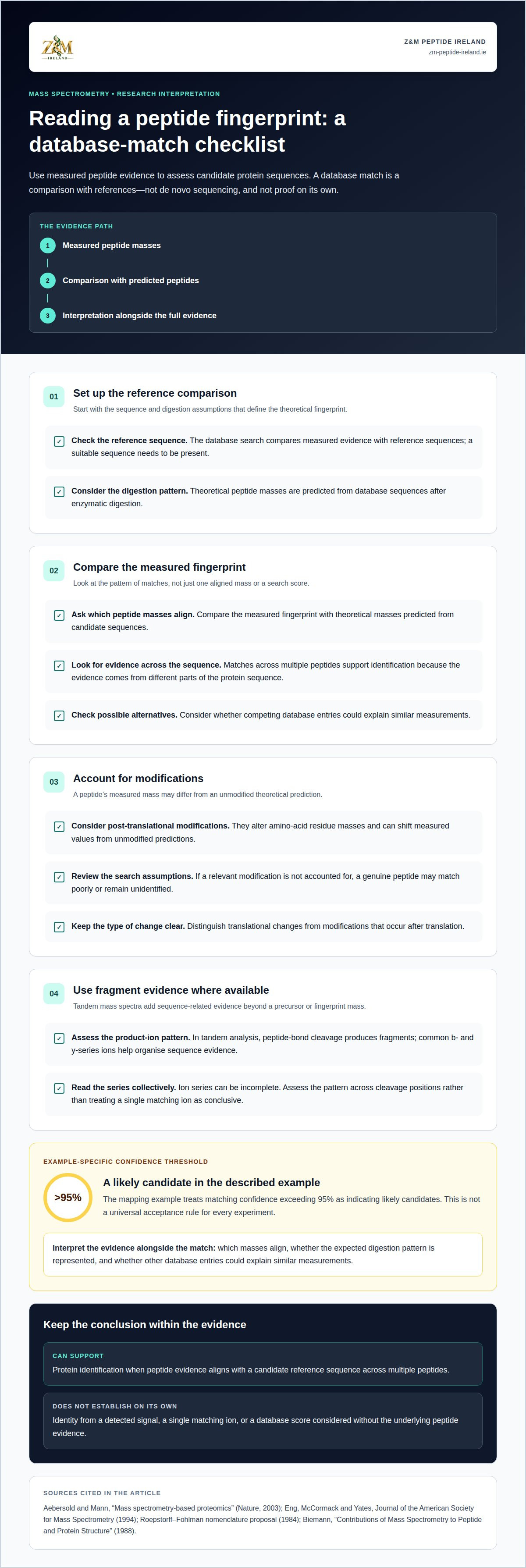
Process peptide spectra by deriving measured peptide masses and comparing them with theoretical masses predicted from database sequences after enzymatic digestion. Evaluate candidate matches alongside the digestion pattern and possible modifications, rather than treating a database score as proof on its own.
In peptide mapping, a protein database provides reference sequences from which theoretical peptide fingerprints are generated. The search compares these predicted masses with the measured fingerprint. Matches across multiple peptides support protein identification because the evidence comes from several parts of the sequence. This approach depends on a suitable reference sequence being present in the database.
In the described mapping example, proteins with matching confidence exceeding 95% are considered likely candidates for the target protein. That threshold indicates a likely identification in that example, not a universal acceptance rule for every experiment. A strong match should be read alongside the peptide evidence: which masses align, whether the expected digestion pattern is represented, and whether competing database entries could explain similar measurements.
Modifications can complicate this comparison. Post-translational modifications alter amino-acid residue masses, shifting measured values from unmodified theoretical predictions. If a search does not account for a relevant modification, a genuine peptide may match poorly or remain unidentified. When interpreting candidate sequences, distinguish translational changes from modifications that occur after translation.
Database matching differs from de novo sequencing: matching requires a reference sequence, while de novo methods can infer sequences without one. This distinction clarifies what a laboratory result establishes and what it depends on. A peptide fingerprint can support protein identification, but identification alone does not quantify sample purity. Purity interpretation requires evidence about the measured components and analytical method, not simply a candidate name or confidence score.
Bottom-up mass spectrometry workflow
A bottom-up experiment turns a protein sample into measurable evidence through linked stages. The result depends on how sample preparation, separation, instrument measurements and database interpretation work together. Each stage shapes what the next can establish about the molecule.
First, enzymatic digestion breaks the protein into peptides. The resulting mixture represents parts of the original sequence, not the intact protein alone. Incomplete digestion can change which peptides appear, while modifications can alter their measured masses. Both factors affect how closely the observed evidence corresponds to expected peptides.
Next, liquid chromatography separates peptides over time before they enter the mass spectrometer. The instrument records mass-to-charge measurements as signals during separation. In tandem analysis, selected peptide ions are fragmented to produce further measurements for sequence interpretation. This connects a chromatographic signal to peptide-level evidence, rather than treating every signal as an independent protein identification.
Data processing groups related signals and compares measured peptide evidence with theoretical predictions from protein databases. Interpretation depends on the selected database, digestion assumptions and modifications included in the search. A candidate supported by several consistent peptide matches provides a stronger basis for identification than one supported by an isolated mass. The match still depends on the reference sequences available to the analysis.
The practical takeaway is to read the output as a chain of evidence. A database match can identify a likely protein, but it does not by itself establish the purity of the original sample. Purity interpretation also depends on whether signals from other components were resolved and reported, and on the method’s ability to detect them. For research decisions, distinguish protein identification, peptide sequence evidence and sample composition. These related outputs answer different analytical questions.
Z&M Peptide Ireland: Research Peptides
This section does not confirm that Z&M Peptide Ireland currently supplies Research Peptides. Researchers considering a procurement enquiry should verify current product information directly with the business and check that any material meets the requirements of the planned experimental programme.
Use the workflow above to clarify the research question and the evidence an analysis needs to provide. A database match can support protein identification, but it does not establish the purity of a supplied material. Keep procurement and analytical decisions distinct, and assess research materials against the requirements of the planned laboratory work.
Verify product details and documentation before making a procurement decision.
Plan the next analytical decision
Before starting a peptide study, define what evidence will support the intended research decision. Sequence confirmation calls for evidence that supports residue assignment; a composition question needs an approach that can distinguish relevant components. Recording this distinction in the study plan helps align the method, sample preparation and interpretation criteria.
For peptide mass spectrometry analysis, treat the analytical question as part of experimental design, not an afterthought. Specify what the result must establish, how modifications could affect interpretation and which limitations matter to the decision. This gives researchers a clearer basis for discussing results in their experimental context, without treating one identification as a complete assessment of every sample attribute.
For research peptide procurement, visit Z&M Peptide Ireland to review the information available for your research needs.
Z&M Peptide Ireland and Research Peptides
Z&M Peptide Ireland supplies Research Peptides, a relevant next step for readers exploring peptide mass spectrometry analysis.
Turn to Z&M Peptide Ireland when your research moves from understanding analytical methods to sourcing Research Peptides for your work.
Frequently Asked Questions
What is peptide mass spectrometry analysis and how does it work?
Peptide mass spectrometry analysis measures peptide ions to support identification and characterisation. It can address different questions: whether a peptide’s measured mass fits an expected molecule, and whether its fragments support a proposed sequence. These are distinct conclusions. For example, a mass match alone may narrow the candidates, while fragment evidence can add information about residue order.
What sample preparation methods are used for peptide mass spectrometry?
Preparation depends on whether the starting material is an isolated peptide, a protein or a complex mixture. Protein samples may be digested enzymatically to generate peptides for mapping; the enzyme and digestion conditions influence which fragments are produced. Researchers also consider sample cleanliness and compatibility with the chosen instrument. Unwanted components can interfere with ion signals, so preparation should suit both the sample matrix and the analytical objective.
What are the steps in an LC-MS/MS peptide analysis workflow?
An LC-MS/MS workflow generally includes sample preparation, chromatographic separation, mass measurement, precursor selection, fragmentation and data interpretation. The instrument records peptide signals during separation, then analyses selected ions and their fragments. Processing software relates these measurements to candidate sequences. For practical interpretation, record the sample type, digestion approach and database-search assumptions alongside the output. These details help explain why a peptide was included, excluded or assigned to a candidate.
How is peptide sequence and identity determined from mass spectra?
Sequence and identity are inferred by evaluating precursor-ion measurements alongside fragment-ion patterns. Differences between fragment masses can indicate amino-acid residues, while the overall pattern is compared with candidate sequences. A reference database match supports identification only when the observed evidence fits the proposed peptide. Modifications can shift expected masses, so an unexplained mismatch may reflect altered chemistry rather than a different sequence. De novo interpretation does not require a reference sequence.
How accurate and sensitive is peptide mass spectrometry analysis?
The fact list states that current mass spectrometers can detect analytes at attomolar concentrations (10⁻¹⁸). In the described peptide-mapping example, matches with confidence exceeding 95% are considered likely candidates, but this is not a universal accuracy threshold; results depend on the peptide evidence, sample and method.
